
Plant Anatomy: Lamellar Collenchyma, Sclereids and Fibres (Part 5)
Explore “Lamellar Collenchyma, Sclereids and Fibres” in this Plant Anatomy lesson. Part 5 of the five-part series brings together the source material and lesson topics. Use the contents links to find a section, read the notes, and consult the complete original teaching slides below.
Lesson topics
- Lamellar collenchyma
- Sclerenchyma
- SCLEREIDS | FIBRES
Contents
REQUEST THE COMPLETE DOCUMENT ON WHATSAPP
Read the original source document
Plant Anatomy – Part 1 (English): original teaching slides for this study series.
Lamellar collenchyma
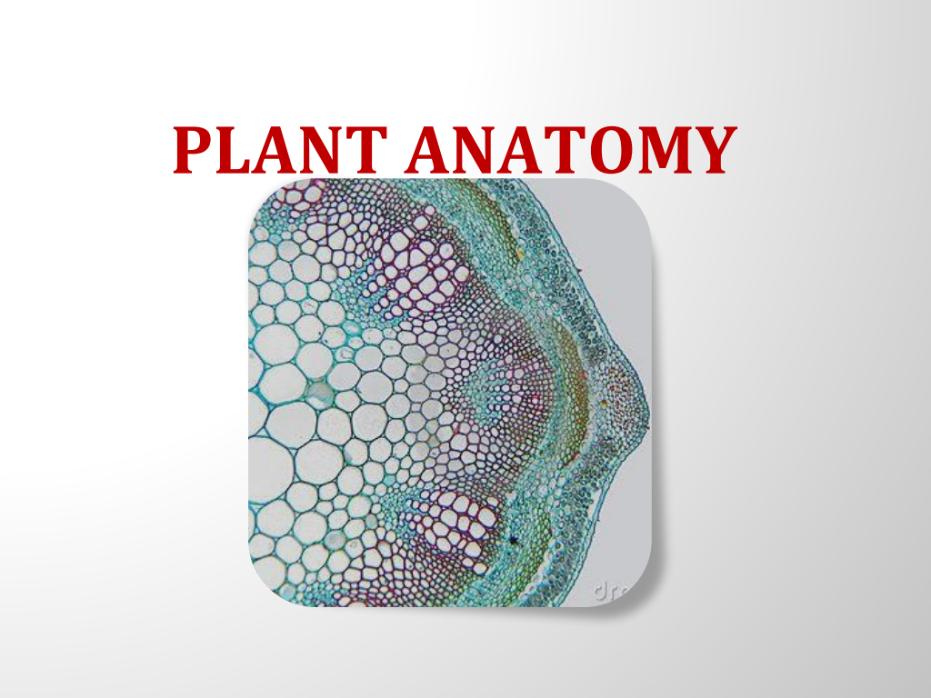
Lamellar collenchyma The collenchyma cells are arranged compactly in layers(rows). The Cell wall is thickening is at tangential walls. These thickening appear as successive tangential layers. Eg: Hypodermis of Helianthus
Sclerenchyma
Sclerenchyma The sclerenchyma is a dead cell and lacks protoplasm. The cells are long or short, narrow thick walled and lignified secondary walls. The cell walls of these cells are uniformly and strongly thickened. The sclerenchymatous cells are of two types: Sclereids
SCLEREIDS | FIBRES
SCLEREIDS | FIBRES Short cells | Long cells Usually short and broad | Narrow, Elongated pointed ends Occurs individually or in small groups | Occurs in bundles Maybe branched | Commonly unbranched Develops from secondary sclerosis parenchyma cells | Derived directly from meristematic tissue
S. DINESH BABU M.Sc.,M.Ed.,M.Phil.,SET.,
S. DINESH BABU M.Sc.,M.Ed.,M.Phil.,SET.,
Review questions and answers
What are the key points about Lamellar collenchyma?
Lamellar collenchyma The collenchyma cells are arranged compactly in layers(rows). The Cell wall is thickening is at tangential walls. These thickening appear as successive tangential layers. Eg: Hypodermis of Helianthus
What are the key points about Sclerenchyma?
Sclerenchyma The sclerenchyma is a dead cell and lacks protoplasm. The cells are long or short, narrow thick walled and lignified secondary walls. The cell walls of these cells are uniformly and strongly thickened. The sclerenchymatous cells are of two types: Sclereids
What are the key points about SCLEREIDS | FIBRES?
SCLEREIDS | FIBRES Short cells | Long cells Usually short and broad | Narrow, Elongated pointed ends Occurs individually or in small groups | Occurs in bundles Maybe branched | Commonly unbranched Develops from secondary sclerosis parenchyma cells | Derived directly from meristematic tissue
More lessons from this document
- Plant Anatomy – Part 1 (English) — Part 1
- Plant Anatomy – Part 1 (English) — Part 2
- Plant Anatomy – Part 1 (English) — Part 3
- Plant Anatomy – Part 1 (English) — Part 4
- Plant Anatomy – Part 1 (English) — Part 5
Conclusion
The important points in Lamellar collenchyma should be reviewed alongside the definitions, examples and comparisons given above. Continue with the other four articles to cover the rest of the source material.